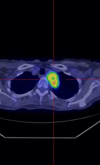
Multimodality
Comprehensive Oncology Staging with Oasis: Advanced Multi-Modality Imaging, Fusion Capabilities, and Precise Tumor Measurement for Accurate Metastasis Evaluation
Unlock powerful tools for staging various types of cancer with Oasis. Utilize multi-modality imaging, fusion options, and precise tumor measurement to accurately assess the extent of metastasis and guide treatment decisions.
Multimodality
Discover the power of seamless fusion with our advanced nuclear medicine applications. With Oasis software, you can effortlessly combine studies from different modalities—like an MRI scan and a PET-CT—without the need for an expensive multimodality camera. Oasis makes fusing images from standalone devices flawless, streamlining your workflow and enhancing diagnostic precision.
MI Register
MIRegister is the next-generation tool designed to elevate your imaging capabilities by enabling precise co-registration of two volumetric datasets, delivering high-accuracy fusion displays for better-informed diagnostic decisions.
What is MIRegister?
MIRegister leverages Mutual Information (MI)—a sophisticated algorithm that analyzes and aligns a volumetric dataset with a reference volume to achieve exceptional precision in image alignment. This ensures that complex multi-modality data is accurately co-registered, providing a cohesive view that is essential for nuanced diagnostics.
With MIRegister's intuitive, user-friendly interface, clinicians can navigate the co-registration process seamlessly, allowing them to spend less time on technical steps and more time on critical clinical decision-making. Additionally, MIRegister's enhanced visualization tools provide detailed, multi-dimensional insights, supporting improved diagnostic accuracy and more comprehensive patient evaluations.
Dual Modality Fusion: Supports combinations such as:
Two SPECT or PET volumes
SPECT/MRI
SPECT/CT
PET/MRI
PET/CT

PET-CT-MR
Elevate your diagnostic capabilities with the Oasis PET/CT package—an essential tool for reviewing volumetric multi-modality data. Fully compatible with data from all major vendors, this unique package makes reading PET studies effortless. With automatic co-registration using advanced Mutual Information Algorithms or the option for manual registration across PET, CT, MR, and/or SPECT images.

Why Oasis V3 for Oncology studies?
Key Features
-
Standardized Uptake Value (SUV) provided in Minimum, Maximum, Average, Peak, and Total Lesion Glycolysis (TLG) values, calculated by four popular methods including Body Weight, Lean Body Mass (James (LBM)), Lean Body Mass (Jamahasatian (LBM - Janma)), or Body Surface Area.
-
Powerful quantification options to manage disease and track lesions over time including all SUV values mentioned above, SUV Max, % change, and metabolic and anatomic volumes.
-
Provides innovative response assessments such as PERCIST.
-
Can compare PET and SPECT datasets
-
Dedicated lesion, linking, and registration managers.
-
Keyboard shortcuts (hotkeys) for tool functions and windowing settings.
-
User definable CT window presents and image layouts with multi-monitor support.
-
Multi-study comparison and auto-linking (up to 4 studies).
-
QPET calculation.
-
Crosshairs bound across all views, fully triangulated views including MIP views.
-
Fusion display with alpha blending.
-
Easily save fused images back to the server and PACS.
-
Save sessions, VOIs, rulers.. and much more back to PACS.
-
Save results and images in JPEG/BMP format.
-
Support for advanced workflows such as "Head and Neck" and "Melanoma."
-
Real time image scrolling.
-
Easily customizable.
-
Full resolution whole body display

SPECT-CT-MR
Maximize your clinical capabilities with our versatile SPECT/CT/MR applications. Load and view a variety of studies, including SPECT reconstruction, CT, MRI, and whole-body scans. Our platform offers powerful tools for enhanced diagnostic accuracy, with features like multi-planar reformation projections, Maximum Intensity Projections (MIP), and fused imaging for deeper anatomical insights.
Dynamic visualization tools, such as cine and splash pages, allow for interactive analysis, while dedicated SPECT pages focus on key orthogonal projections. Customize settings for scaling, color, and layouts to fit your protocol. Plus, compare up to four SPECT/CT scans simultaneously to streamline your workflow and elevate your diagnostic process.

Key Features
-
Automatic Registration - Streamline your workflow for enhanced efficiency.
-
3D Region Analysis - Based on iso-contouring for deeper insights into complex structures.
-
Dedicated Managers - For lesion linking and registration.
-
Study Comparison Features -Facilitate effective follow-up evaluations.
-
Manual Registration Adjustments - Make precise translations and rotations.
-
User-Definable CT Window Presets - Customize your workflow for quick adjustments.
-
Distance Weight Maximum Intensity Projections (M.I.P.) - Enhance visibility of critical areas.
-
Fusion Display Mode - Utilize variable alpha blending and thresholding for seamless visualization of combined datasets.
-
User-Definable Hotkeys - Streamline access to commonly used functions.
-
Standard Orthogonal Slice Displays: Ensure clarity and detail for visualizing intricate anatomical structures.
Lymphography
Experience comprehensive lymphatic imaging with our cutting-edge Lymphography tool! Effortlessly visualize static, dynamic, and whole-body scans to pinpoint lymph drainage basins and sentinel nodes with unmatched precision.
Lymphography empowers you to accurately identify and count sentinel nodes, distinguish them from surrounding nodes, and even discover sentinel nodes in unexpected locations. With the ability to mark nodes for precise biopsy guidance, this tool elevates your diagnostic capabilities. Plus, our advanced Lymphatic Transport Index algorithm ensures reliable, data-driven decisions by calculating precise regions of interest (ROI).

Quantification of Lymphatic Transport Index: Gain valuable insights with our percentage calculation of lymphatic transport, enhancing your diagnostic capabilities.
Bone Scan
Unlock Insights into Skeletal Health!
The Bone Scan application—a vital tool for detecting abnormal skeletal radiopharmaceutical uptake, which indicates increased bone metabolic activity. While primarily used to identify cancer metastasis to the bones, Bone Scans also evaluate conditions such as Paget’s disease, stress fractures, and infections.
Our intuitive processing includes optional analysis, such as drawing rectangular regions of interest (ROI) over the sacroiliac joints, allowing for precise calculation of average counts per pixel. This thorough analysis provides you with essential data for effective diagnosis.

Key Features
-
Sacroiliac (SI) Joint Ratio: Analyze the sacroiliac joint with precision using our easy-to-use ratio calculation.
-
Enhanced Visualization: Mask bladder and “hot spot” activity to improve clarity and focus on bone structures.


